In our February newsletter to our veterinary colleagues we set a 'What's your approach and diagnosis' question and here is the answer and Dr Ursula's advice on treatment for these cases
What we did:
Basic tests in both eyes should include a Schirmer tear test reading, Fluorescein test and intraocular pressure test. The opposite eye will require careful examination as to detect any abnormal changes.
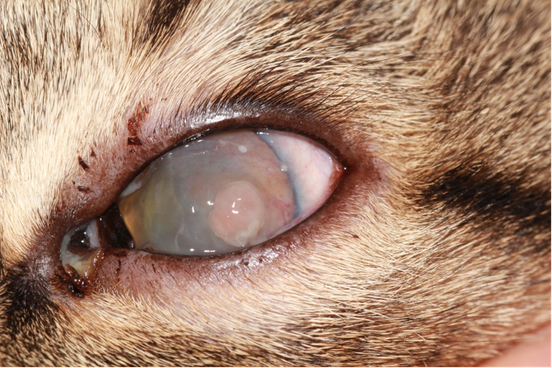
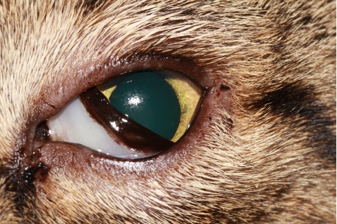
The advanced vascularisation and corneal oedema are consistent with Keratits. There is no fluorescein stain uptake noticed in the cornea. Possible underlying causes and differential diagnosis include:
The following tests can be considered:
Diagnosis: Cytologic examination of the corneal scraping revealed the presence of corneal epithelial cells, neutrophils and eosinophils. Based on those findings the established diagnosis is Eosinophilic keratitis.
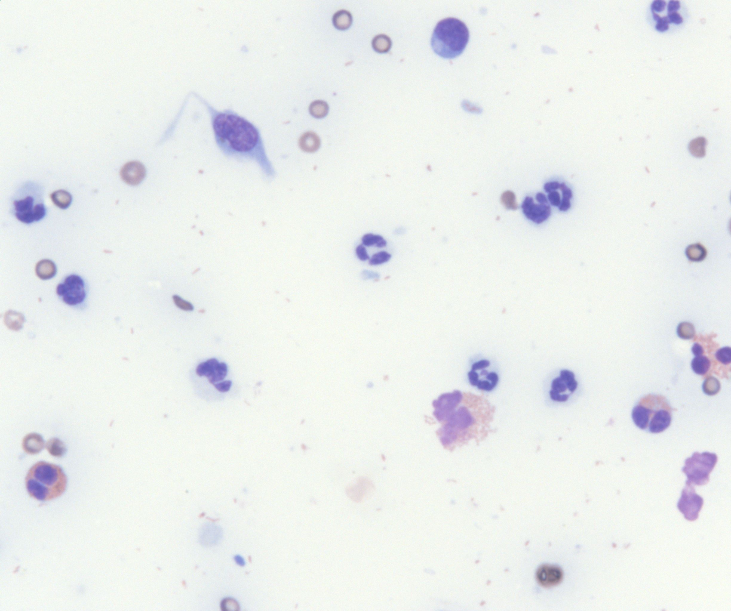
Cytology shows neutrophils, epithelial cells and eosinophils
Eosinophilic keratitis, a condition seen in domestic cats, is characterized by proliferative lesions on the cornea; it is distinct from the dermatologic eosinophilic complex in cats. The exact cause is unknown but it is believed to be an immune-mediated corneal condition in cats. It may affect one or both eyes and it is usually observed in young to middle-aged cats. Typical lesions involve raised, multifocal, plaque-like pinkish-whitish lesions with various degree of vascular ingrowth. The lesions usually start at the lateral or nasal limbus and untreated may progress to involve the entire cornea. The adjacent bulbar conjunctiva and nictitans might be affected as well. There is a distinct form of eosinophilic conjunctivitis with similar appearance, but not affecting the cornea. Eosinophilic keratitis/conjunctivitis may affect one or both eyes and it is usually observed in young to middle-aged cats.
Cytology of the lesions obtained through a corneal/conjunctival scraping reveal the inflammatory process with neutrophils, mast cells, plasma cells, lymphocytes and most typically eosinophils. The detection of eosinophils is diagnostic for this condition.
Eosinophilic keratitis/conjunctivitis usually responds quite dramatically to the application of topical corticosteroids, which are given 2-3 times daily initially to effect and slowly tapered once remission occurs. A combined therapy with topical cyclosporine (0.2% Optimmune) is recommended and will be continued after discontinuation of topical steroids for long-term control of the condition. The treatment with topical cyclosporine can be tapered over time, but recurrence of the condition is possible and the eye requires careful long-term monitoring.
Our treatment plan:
Initial treatment with Maxitrol (Dexamethasone/Neomycin/Polymyxin B) eye drops every 12 hours and 0.2% Optimmune ointment every 12 hours. There was dramatic improvement noticed during the first 2 months of treatment and Maxitrol was reduced to once daily and then discontinued after 3 months of treatment. Topical cyclosporine (Optimmune) was continued as ongoing treatment every 12 hours.
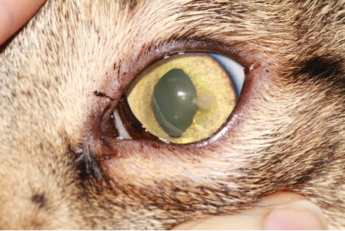
2 months later
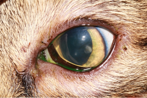
3 months later
6 months later
Treatment led to complete resolution of the corneal lesions and vascularization in the cornea.