
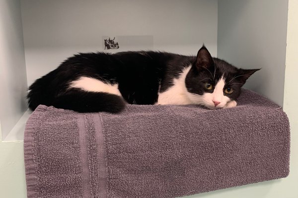
Ava is a beautiful and affectionate 1 year old, she was rescued off the streets of Turkey when she was 6 months old. She travelled to London via Barcelona with her fantastic new family.
She came to us she had been treated for ear mites previously but it now looked like she had an ear infection.
Ava is extraordinarily friendly and let Dr Jeremy examine her ears without even the slightest protest. Her right ear canal was fine but her left ear canal was filled with purulent (pus) discharge which meant we could not visualise the tympanic membrane (ear drum) at all.
We needed to see what bacteria we were dealing with so we could treat with the appropriate antibiotics so we took swabs and submitted these for culture at our laboratory. Dr Jeremy started her on some broad-spectrum antibiotics while we waited for the results.
The results came back showing 2 different types of bacteria but thankfully both of these were sensitive to the particular antibiotic that Ava was taking.
At home Ava’s human family flushed her ears with sterile saline 1-2 times a day. With the cleaning and the antibiotics her ear improved by 75%. Ava was due to be neutered but we wanted her to be 100% before we booked this procedure so we continued her treatment for an additional 10 days.
Ava came in for a revisit and while the infection had cleared it had revealed the presence of abnormal tissue within in the ear canal that was obstructing the ear canal right down by the eardrum. This required further investigation.
Under general anaesthesia we took x-rays of her head and inner ears which showed evidence of infection or fluid/tissue in the left inner ear and a mass in the horizontal ear canal.
For a really detailed examination of the ear drum and investigation of the mass we needed to use our video-otoscope. Video-otoscopy uses a very small fibreoptic light source and camera that we can magnify greatly to provide a close-up and hugely detailed picture of the area of interest.
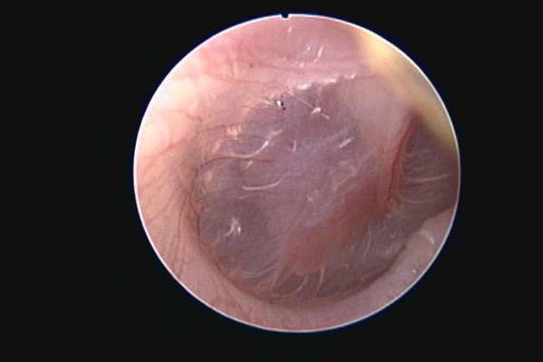
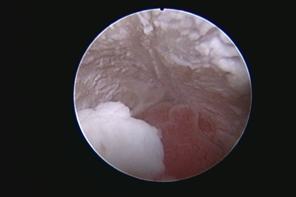
When we examined Ava’s ear we saw that the tissue was actually a 5 x 5mm growth (see image 1, image 2 is her healthy ear) that was projecting out into the canal on a stalk (polyp) which Dr Jeremy removed with gentle traction pressure. Due to the evidence of fluid accumulation in the inner ear a procedure called a myringotomy was performed. A myringotomy involves piercing the ear drum in a precise location with a very small sharp catheter to then collect fluid which is trapped on the other side and gently flush any infection out.
Ava recovered brilliantly from her procedure and was back to affectionate adorable self immediately post-operatively.
The mass came back as a benign nasopharyngeal polyp. These polyps arise from the inner ear and can make their way into the back of the throat or the ear as in Ava’s case.
The infection in her inner ear was treated for 6 weeks with the appropriate antibiotics and Ava has never looked back.
Ava has had such a journey, and she still greets everyone with big purrs and cuddles!